For every heartbeat, the heart sends out more than an electrical signal. It also creates a magnetic field so faint that detecting it outside the body is a formidable measurement challenge. In the experiment, the researchers brought a tiny diamond sensor to within about a centimeter of the chest and began collecting those barely perceptible signals. Heartbeat after heartbeat, the measurements accumulated until the noise gave way to a recognizable pattern of cardiac activity.
Every heartbeat involves electrical activity that moves through the heart muscle. That activity can be measured electrically with an electrocardiogram, or ECG, but it also produces a tiny magnetic field that can be detected outside the body.
That second signal is known as a magnetocardiogram, or MCG. Unlike an ECG, it does not require electrodes touching the skin, and magnetic fields are much less affected by the electrical conductivity of tissues such as fat, fluid and lungs. MCG can therefore provide information that complements an ECG, including information that can help localize cardiac electrical activity.
The difficulty is that the magnetic signal from the heart is extremely weak. Environmental magnetic noise can overwhelm it, which is why conventional MCG systems have commonly depended on highly sensitive sensors and stringent magnetic shielding.
The new work takes a different approach. Three groups independently developed nitrogen-vacancy, or NV, diamond magnetometers and used them to detect magnetic signals from a human heart. The sensors operate at room temperature and use the quantum properties of defects in diamond to measure magnetic fields.
The experiments were performed under very different magnetic conditions. One sensor operated inside a multilayer magnetically shielded room with no static magnetic bias. A second operated in a partially shielded environment with a roughly 1-millitesla bias field. A third was used in an unshielded environment, also with a roughly 1-millitesla bias field.
Together, the experiments tested not only whether a diamond sensor could detect human cardiac magnetism, but also how such sensors behave when the magnetic environment becomes progressively less controlled.
A diamond sensor sees the heartbeat
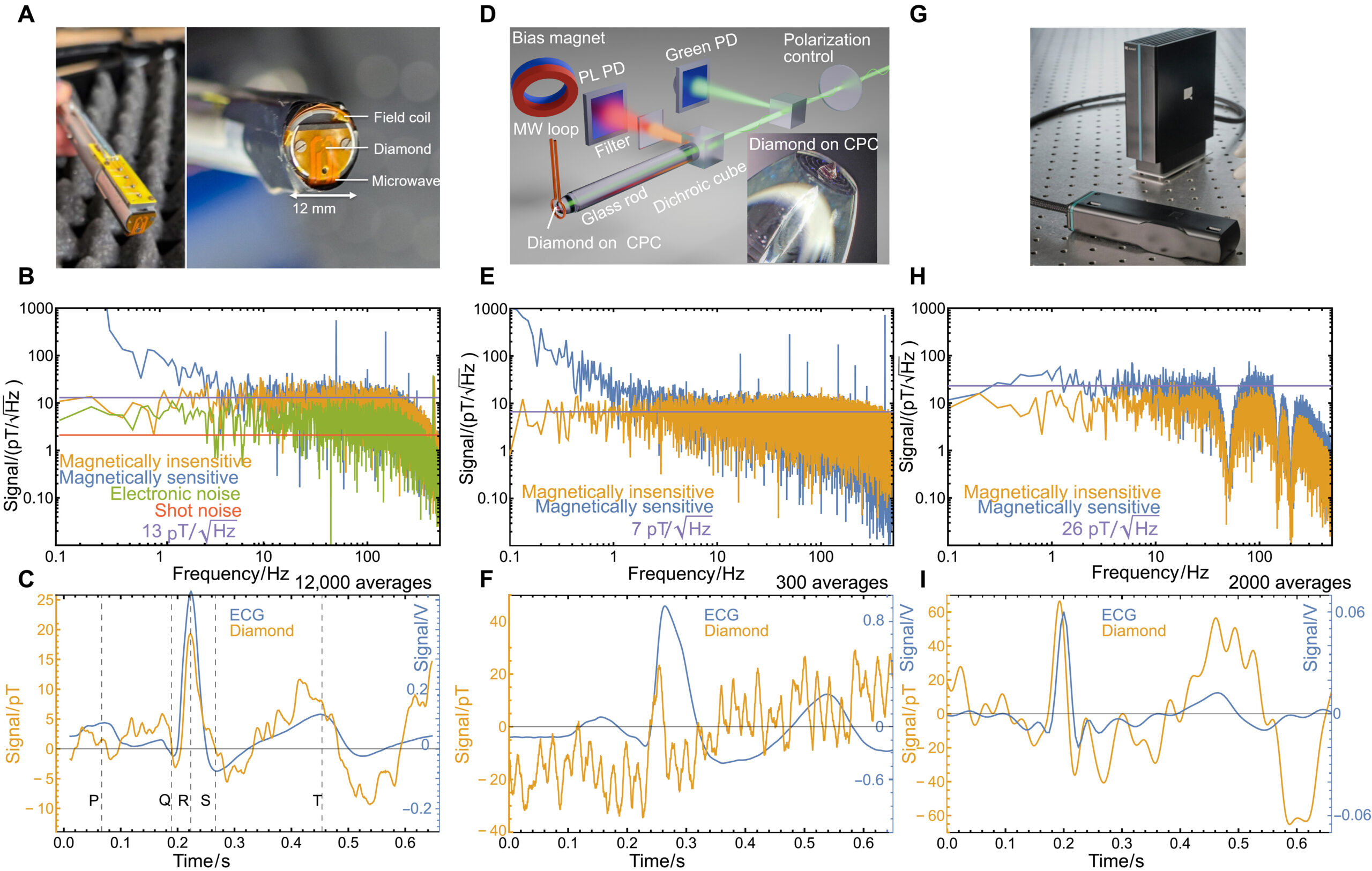
The first measurements were made at the Helmholtz Institute Mainz using a fiberized NV magnetometer designed as a transportable, endoscope-like sensor.
Its diamond was only 0.5 by 0.5 millimeters across and 0.18 millimeters high. The sensor operated without a static magnetic bias field and achieved a magnetic sensitivity of 13 picotesla per square root hertz between 20 and 30 hertz.
During measurements, the sensor was positioned about 1 centimeter from the chest of a seated person. An ECG was recorded at the same time and used to identify individual heartbeats so that the magnetic signals from many cardiac cycles could be aligned and averaged.
That averaging was essential. The magnetic signal was not visible in the diamond sensor trace in real time. Instead, the researchers accumulated repeated heartbeats until the small, consistently timed cardiac signal emerged from the noise.
For the displayed Mainz measurement, the MCG trace represented 12,000 averaged cardiac cycles. The resulting signal showed the familiar sequence associated with cardiac electrical activity, including the P wave, QRS complex and T wave.
The other two diamond systems also produced recognizable MCG traces. The ZAQuant sensor had a measured sensitivity of 7 picotesla per square root hertz, while the Q.ANT sensor had a sensitivity of 26 picotesla per square root hertz. Their displayed cardiac traces were based on 300 and 2,000 averaged heartbeats, respectively.
The different numbers of averages do not directly indicate that one sensor was intrinsically better than another. The researchers emphasize that the quality of the averaged traces depended strongly on the magnetic environment, including correlated low-frequency noise that is not efficiently removed by simply averaging more measurements.
Three sensor designs, three magnetic environments
The three magnetometers were built differently.
The Mainz device used the smallest diamond, measuring 0.5 by 0.5 by 0.18 millimeters, inside a compact 12-millimeter-diameter sensor head. It used zero static magnetic bias and magnetic-field modulation.
The ZAQuant system used a 0.5-by-0.5-by-0.5-millimeter diamond and operated with a roughly 1-millitesla bias field. The Q.ANT device contained a larger 1-by-1-by-0.5-millimeter diamond and also operated at about 1 millitesla.
Their reported sensitivities were 13, 7 and 26 picotesla per square root hertz, respectively. The systems also differed in their optical designs, microwave control, readout methods, sensor-head integration and operating environments.
Those details matter because a larger diamond does not automatically produce a lower noise floor. The overall sensitivity reflects the performance of the complete magnetometer, including optical collection, spin-resonance contrast, microwave uniformity, technical noise and stability against temperature, mechanical and magnetic-field changes.
At Mainz, the diamond was operated at zero magnetic bias. A 6.12-kilohertz modulated magnetic field was applied along the diamond’s [100] crystallographic direction, allowing the researchers to recover magnetic sensitivity from NV centers along the crystal axes without using a static bias field. The microwave system was centered at 2.87 gigahertz and was modulated and actively locked to resonance.
The Q.ANT system used a 1-by-1-by-0.5-millimeter diamond containing about 1.7 parts per million negatively charged NV centers. Its fluorescence was collected with a balanced photodiode system, while a roughly 1-millitesla magnetic field was supplied by a Halbach ring. The integrated optical design had an estimated fluorescence-collection efficiency of about 40 percent based on optical simulations.
Checking the diamond signal against another sensor
Detecting a waveform after extensive averaging is not by itself enough to establish that the diamond system is measuring the heart’s magnetic field rather than an electrical signal leaking into the electronics.
The researchers therefore performed additional validation.
They measured the cardiac magnetic field over the chest using a 49-channel array of optically pumped magnetometers, or OPMs, in a magnetically shielded room. The OPM array had a nominal sensitivity of about 15 femtotesla per square root hertz, considerably below the noise levels of the diamond sensors used in the study. The array’s sensors were spaced 2.5 centimeters apart and positioned at least 5 centimeters from the chest.
The OPM measurements were visible in real time and were averaged over 100 heartbeats after filtering between 3 and 30 hertz.
The researchers then used the OPM map to identify two positions, P1 and P2, for measurements with the diamond sensor. At P1, they averaged 12,000 cardiac cycles, while P2 used 6,000. The different averaging counts reflected the available recording time and the specific purpose of each measurement.
The resulting diamond and OPM measurements produced comparable MCG peak amplitudes. The diamond measurement also changed polarity between the two positions as expected from the spatial variation of the magnetic field.
The researchers separately tested whether the ECG could have been picked up electronically by the diamond system. They recorded the diamond signal at two positions where the MCG peak had different polarities while keeping the ECG configuration unchanged. The position-dependent reversal ruled out electronic cross-talk as the explanation for the observed signal.
The two types of sensors did not produce identical waveforms. Their R-peak amplitudes happened to match, but the T-wave shapes differed noticeably.
The researchers attribute this difference to the sensors’ different positions, sensing volumes and sensitive-axis orientations. The OPM array itself showed substantial variation in T-wave morphology from one sensor position to another. Because the magnetic field varies across the chest, two sensors looking at the same heart from different locations or directions can record different versions of the cardiac waveform.
The harder test is outside the shielded room
Detecting an MCG signal in a carefully shielded environment is an important demonstration, but real-world environments contain magnetic interference from many sources.
The researchers therefore explored gradiometry, a technique in which two spatially separated sensors measure the magnetic field simultaneously and their signals are compared.
The idea is straightforward. A distant environmental magnetic field can affect both sensors similarly, while a localized cardiac field changes more strongly between the two positions. Subtracting the two measurements can therefore suppress common magnetic interference while retaining spatially local signals.
At ZAQuant, two identical NV sensor heads were placed 78 millimeters apart inside a single-layer magnetically shielded room. Helmholtz coils were used to apply controlled background fields representing environmental interference, while localized magnetic signals were introduced to imitate biomagnetic sources.
The applied test fields were several hundred nanotesla. Simply subtracting the measurements from the two sensors substantially reduced the common field and the resulting noise. At low millihertz frequencies, the individual sensors had noise spectral densities greater than 100 nanotesla per square root hertz. The gradiometric signal reduced that noise by almost two orders of magnitude.
There was, however, a limit. The gradiometer’s linear range was constrained by the approximately 200-kilohertz magnetic-resonance linewidth of the ZAQuant sensor, with suppression degrading beyond about ±500 nanotesla.
The authors note that active frequency tracking and closed-loop feedback could extend this range to field variations of several microtesla. They also point to the scalability of the diamond platform, which could allow gradiometric sensor separations ranging from millimeters to meters depending on the application.
The experiment also exposed a more complicated problem. Environmental magnetic noise is not always identical at the two sensors. Differences in sensor gain, orientation, local shielding and the geometry of the noise source can cause the same disturbance to appear differently in the two channels.
In addition, real-world interference can change over time and overlap with the frequency range of cardiac signals. Under those conditions, simply subtracting one sensor from the other is not necessarily sufficient.
Separating a weak heartbeat from changing noise
To explore that more difficult situation, the researchers tested a blind source separation, or BSS, approach using simulated two-channel NV-magnetometer data.
The simulation contained a weak cardiac-like magnetic signal and a much stronger environmental magnetic-noise component whose amplitude and frequency changed over time. The two simulated sensors received different mixtures of the two sources.
Before separation, the simulated sensor channels were dominated by the environmental disturbance, and the cardiac component was not directly apparent. Applying BSS separated the two components, recovering signals corresponding to the cardiac-like source and environmental noise.
The method did not require the researchers to specify the exact waveform of the environmental noise beforehand. Instead, it used the fact that the cardiac and environmental sources couple differently to the two spatially separated sensors and have different temporal or statistical characteristics.
This was a simulation rather than a demonstration of real-time human MCG under uncontrolled conditions. Its role in the study was to illustrate how gradiometry could be extended from simple subtraction into an adaptive source-separation problem when the noise is spatially nonuniform or changes over time.
Real-time cardiac detection remains out of reach
The experiments establish that NV-diamond magnetometers can detect human cardiac magnetic signals, but the measurements also expose the central limitation of the current systems.
The sensors can produce recognizable MCG waveforms after averaging many heartbeats. They cannot yet provide the signal quality needed for routine real-time cardiac measurements.
The Mainz experiment required 12,000 averages, while the other demonstrations also relied on hundreds or thousands of cardiac cycles. The Q.ANT sensor produced an MCG signal in an unshielded environment after filtering, but the study notes that environmental noise remains a major constraint.
The researchers estimate that sensitivities around 10 picotesla per square root hertz are sufficient to detect MCG signals after enough averaging, but reducing the averaging time toward low-average or real-time measurements would require improvement toward roughly 1 picotesla per square root hertz. That represents about an order-of-magnitude improvement over the sensors used in these measurements.
The authors place that target in the context of existing magnetometry. Recent NV systems have achieved sensitivities below 10 picotesla per square root hertz, while subpicotesla-per-square-root-hertz sensitivity is currently reached by SQUID and OPM systems used for biomagnetic measurements.
One limitation comes from the way the diamond sensors are currently operated. The ensemble magnetometers in this work use continuous-wave protocols that are approaching practical sensitivity limits. Photon shot noise is an important contribution, and increasing optical excitation can reduce that noise, but additional laser power is constrained by effects including power broadening, heating and laser-induced decoherence.
Several approaches are discussed in the paper as possible ways to improve sensitivity. These include infrared absorption magnetometry, pulsed sensing with dynamical decoupling, microwave cavity-enhanced detection, and quantum-enhanced approaches such as spin squeezing and quantum gain amplification. The authors describe the more quantum-enhanced strategies as promising but experimentally complex at present.
Another approach is to concentrate magnetic flux before it reaches the diamond. Flux concentrators can collect magnetic flux over a larger region and channel it into the sensing area. The paper cites demonstrations of more than two orders of magnitude signal enhancement using such structures, while noting that collecting flux over a larger area reduces spatial resolution.
For now, the three sensors demonstrate different pieces of the problem rather than a single finished clinical system. The Mainz sensor provided the clearest shielded human MCG measurement. The ZAQuant device demonstrated the gradiometric noise-suppression approach, while the Q.ANT system operated in an unshielded environment. The authors report that the gradiometric sensitivity remained about an order of magnitude worse than the single-sensor performance, and that a substantial sensitivity gap still separates these systems from routine clinical use.
The measurements therefore leave a specific technical challenge: improving the sensitivity of compact diamond magnetometers enough that the heart’s magnetic waveform can be detected with little or no averaging while environmental magnetic interference is simultaneously controlled.
The study was published in Science Advances.