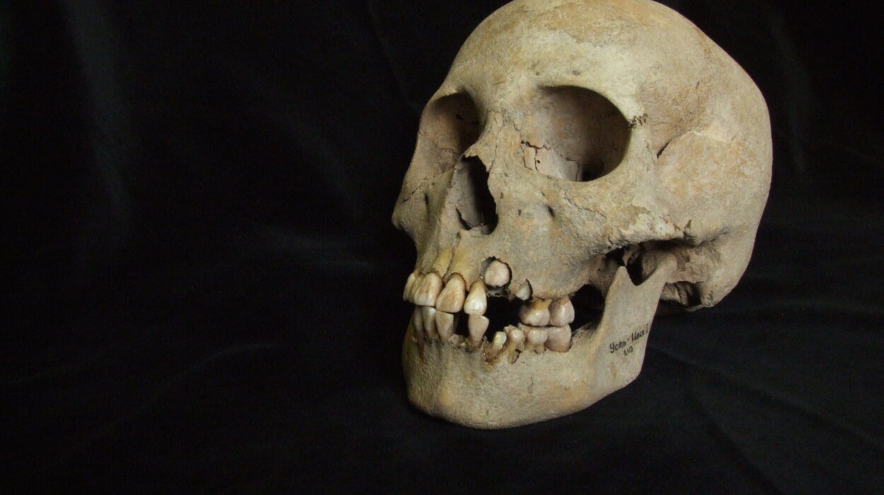
Some 90,000 to 145,000 years ago, a young adult died after spending part of their life with an unusual injury carved across the left side of the lower face, and thousands of years later, high-resolution imaging has allowed scientists to reconstruct what may have happened: a sharp object appears to have grazed a tooth before striking the jaw, leaving behind a wound that had already begun to heal.
When Qafzeh 25 was recovered from Qafzeh Cave in Israel, the skeleton was already badly damaged. Much of it had been broken after death, and earlier excavations had resulted in the loss of much of the lower limbs. The remains had therefore undergone a complicated history between burial and modern study.
Yet the skeleton had one unusual feature that demanded a closer look.
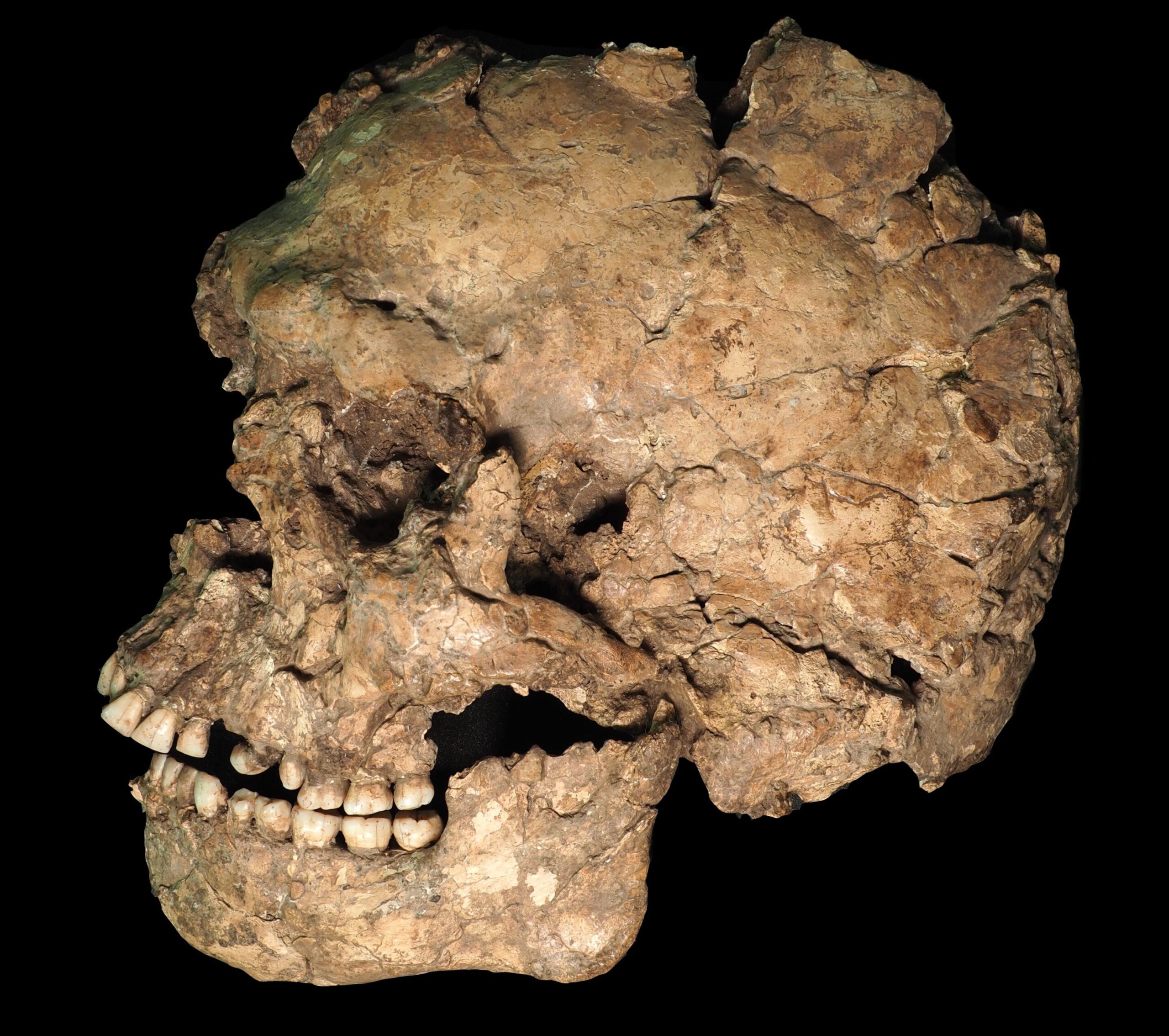
On the left side of the mandible, or lower jaw, researchers identified a narrow linear mark running across the bone and into the left lower third premolar. At first glance, the damaged region was complicated by numerous fractures and other alterations. The researchers therefore examined the original fossil at several levels, combining direct macroscopic and microscopic observations with high-resolution micro-computed tomography, or mCT.
That approach revealed a much more coherent pattern than the broken surface initially suggested. The mark had an overall V-shaped cross-section, extended about 9 millimeters along the cheek-facing surface, and affected roughly 7 millimeters of the tooth itself. Part of the groove crossed the tooth root and then continued into the mandibular bone.
The geometry and trajectory became important because they were not simply the random result of the skeleton’s later deterioration. The researchers concluded that the combination of the mark’s converging shape, position and direction was consistent with an injury produced by a sharp object.
Their reconstruction is strikingly specific. They propose that the object most likely grazed the crown of the premolar before striking the upper border of the mandible, removing some dentine and bone in the process. The later fractures that split and displaced part of the tooth crown, however, were not caused by that original impact. Those fractures followed a broader postmortem fracture pattern affecting nearby teeth.
The distinction matters. The researchers were not simply looking at a broken ancient tooth and calling it a wound. They were separating an injury that happened while the person was alive from damage accumulated long after death.
And the most important evidence came from what happened to the bone afterward.
The wound did not happen at the moment of death
Around the linear mark, the mandibular surface contained localized patches of porosity. Elsewhere, the bone was comparatively smooth and compact. The margins around the affected area were also thickened and projected outward from the normal curvature of the jaw.
Those features are consistent with bone remodeling—the process through which living bone responds to injury. The researchers interpreted the porous areas as evidence of activity associated with healing and the thickened margins as consistent with callus formation during fracture repair.
That changes the story dramatically.
Whatever caused the injury, Qafzeh 25 did not die instantly from it. The injury was antemortem, meaning it occurred during the individual’s life, and the bone had already begun responding to the damage.
The paper therefore concludes that the individual survived for a significant period after the injury. The researchers also note that the trauma exposed dentine but did not extend into the tooth’s pulp chamber, which may help explain why they found no evidence of an associated dental infection.
The exact length of survival cannot be established from the remains. What can be established is that the biological response took place before death.
That turns a small mark on an ancient jaw into a record of something that happened during the individual’s life—and of the body’s attempt to repair it.
The location raises the possibility of a violent encounter
The researchers considered how such an injury might have occurred.
Sharp-force injuries are exceptionally uncommon in the Middle Paleolithic archaeological record. The paper discusses Shanidar 3, a Neanderthal with a rib puncture interpreted as possible sharp-force trauma, as the closest comparable case. Even there, alternative explanations cannot be completely excluded.
Qafzeh 25 presents an additional clue: the injury is on the left side of the face.
The authors note that forensic studies of modern populations have found that craniofacial injuries from blows occur more frequently on the left side, a pattern often attributed to right-handed attackers facing their victims. The researchers emphasize that the side of an injury cannot by itself establish that violence occurred. But when that lateral location is considered alongside a lesion consistent with sharp-force trauma, they argue that an interpersonal explanation becomes more plausible than an accidental one.
That wording is important. The fossil does not preserve the attacker, the weapon, or the circumstances surrounding the event. The researchers therefore do not claim that interpersonal violence has been demonstrated beyond doubt.
An accident remains possible.
But the pattern makes a violent encounter a serious interpretation.
The paper describes Qafzeh 25 as an archaic Homo sapiens individual from the Middle Paleolithic and argues that, if the interpretation is accepted, the specimen would represent the earliest documented case of sharp-force trauma in the archaeological record.
A missing tooth crown adds another unresolved piece
The injury becomes even more intriguing when the researchers traced its apparent trajectory upward toward the upper jaw.
The path of the mandibular mark corresponds spatially with the position of the upper left canine, whose crown is missing. The root remains in place and is covered by sediment. High-resolution imaging showed a horizontal fracture affecting the teeth on the left side of the maxilla, while the canine crown itself is absent.
It would be tempting to connect the missing crown directly to the injury.
The researchers do not.
They found no definitive way to establish that the canine crown was lost during the same event. The exposed root also lacks the smoothing or wear that would be expected if the dentine had remained exposed and functionally used for a prolonged period. That leaves open the possibility that the crown was lost around the time of the injury, but the evidence is insufficient to prove the connection.
That uncertainty is part of the significance of the study. Ancient skeletal injuries rarely come with a complete narrative. Instead, researchers assemble a sequence from the surviving physical traces and distinguish what those traces establish from what they merely make plausible.
The same scan uncovered a hidden problem inside a tooth
The investigation was not limited to the injury.
The researchers used the high-resolution mCT scans to inspect the remaining teeth of Qafzeh 25 for previously undocumented pathology. One of the most revealing findings was hidden inside the left lower fourth premolar.
The tooth contained a small area of demineralized enamel beneath an apparently intact outer surface. The feature measured about 1.5 millimeters in length, 0.25 millimeters in width and 0.35 millimeters in height, with a calculated volume of 0.0513 cubic millimeters. Because the enamel surrounding the cavity had a density lower than healthy enamel but higher than the surrounding sediment and dentine, the researchers interpreted the feature as a hidden carious lesion, sometimes called hidden caries.
This was not an obvious cavity visible on the outside of the tooth.
Instead, the abnormality was enclosed beneath a thin layer of enamel, meaning that conventional inspection could have missed it. The researchers concluded that the internal demineralization resulted from an infectious or bacterial process and identified it as a non-cavitated carious lesion.
The finding expands the dental record of Qafzeh in another way. It is the first documented carious lesion in a premolar within the Qafzeh sample.
With this lesion included, the researchers report that at least 10 carious teeth are now documented from the assemblage. Caries had already been reported in several Qafzeh individuals, so the discovery does not establish that Qafzeh 25 was uniquely affected. Instead, it adds another piece to a broader pattern of oral disease at the site.
Not every damaged tooth was a cavity
The researchers also examined the lower first molars, where enamel loss was visible along the mesiolingual margins of both teeth.
Those defects could have looked similar to carious lesions previously described in another Qafzeh individual. But the new mCT images did not show the enamel demineralization expected from bacterial activity.
Instead, the researchers found smooth, polished edges confined to the enamel and interpreted the defects as enamel chipping that occurred during life, followed by continued tooth function.
This distinction illustrates why the imaging was important. A superficial defect can resemble disease when viewed only from the outside. Examining the internal structure allowed the researchers to distinguish between two different processes: one associated with caries and another more consistent with mechanical damage.
The study also confirmed a pronounced rotation of Qafzeh 25’s left lower fourth premolar. The tooth is rotated by about 65 degrees in a distobuccal direction, while its root and contact with the neighboring premolar remain in their original positions. Because the rotation cannot be attributed to postmortem movement, the researchers regard it as a developmental characteristic rather than damage caused after death.
A similar rotation of the same tooth occurs in Qafzeh 11, although in a different direction. The recurrence of dental anomalies within the Qafzeh sample has previously raised the possibility of hereditary influences, and the authors note that some individuals at the site may have been biologically related.
The skeleton itself preserves evidence of deliberate burial
The injury and dental disease are only part of what Qafzeh 25 can tell researchers.
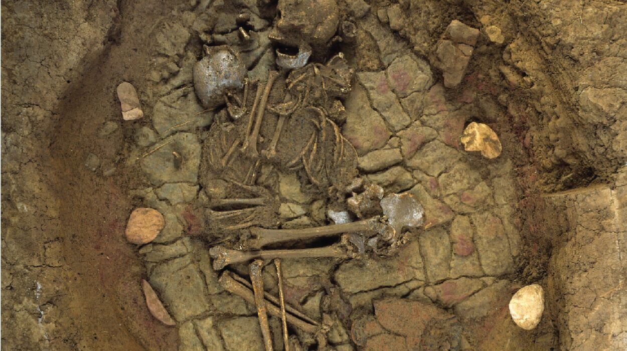
The skeleton was recovered from Layer XVII during the final excavation campaign at Qafzeh Cave in 1979. It was found close to the remains of several immature individuals. Because the skeleton was fragile and extensively altered after death, it was removed in a block of sediment and later excavated under controlled laboratory conditions.
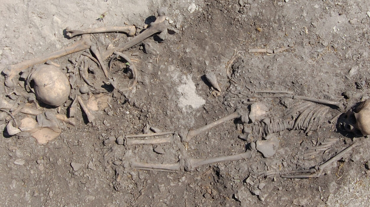
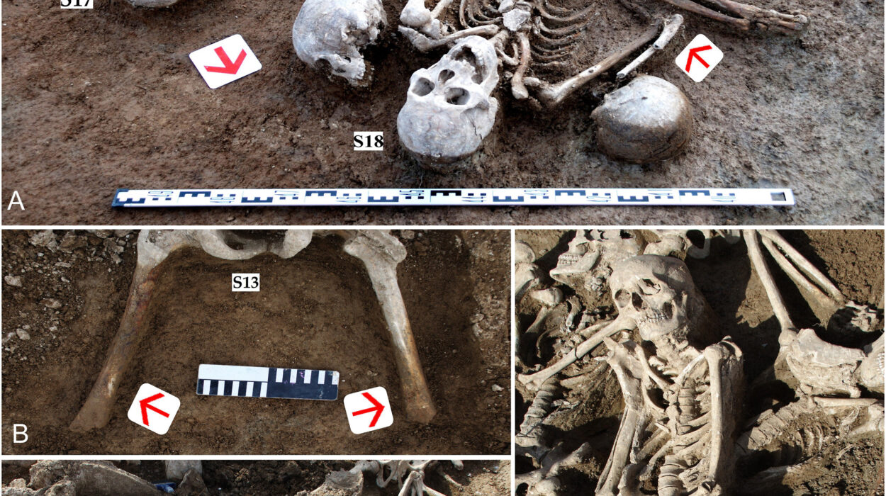
Qafzeh Cave contains a major Middle Paleolithic human assemblage. The site’s Mousterian layers have been dated to approximately 92,000 to 145,000 years ago, and most of the human remains were recovered from Middle Paleolithic layers, particularly Layer XVII. The assemblage includes adults as well as an unusually large number of children and adolescents, many of whom were deliberately buried.
For Qafzeh 25 specifically, there is no directly observed burial pit.
But the skeleton remained in anatomical position. Its bones had not been displaced from the space originally occupied by the body, even though extensive postmortem fracturing had occurred. The researchers interpret that preservation as evidence that the body decomposed within a filled area and conclude that the individual was deliberately buried.
The broader taphonomic evidence reinforces that interpretation.
The skeleton’s fractures were characterized as postmortem, many of them modern, with extensive crushing and collapse of the cortical bone. The fracture surfaces showed the right angles and jagged edges expected from dry-bone breakage. At the same time, the researchers found no signs of weathering, trampling, root etching, or carnivore tooth marks on the examinable surfaces. The bones remained in anatomical association despite their severe compression.
Together, those observations indicate that the remains were buried relatively rapidly in a stable depositional setting and remained largely undisturbed afterward, consistent with other Qafzeh burials.
The burial therefore provides a second behavioral dimension to the story. Qafzeh 25 was not simply an individual who suffered an injury and later became a fossil. The circumstances in which the body was deposited also preserve evidence of how the dead were treated.
A sharper look changed what scientists could see
One reason the new findings were possible is that the researchers were able to look much more deeply into the fossil than earlier investigations could.
A previous reconstruction of the Qafzeh 25 mandible used CT data with voxel dimensions of 540 micrometers in width and height and 625 micrometers in thickness. The researchers in the new study used a resolution 18 times higher, scanning the maxilla and mandible at a 30-micrometer isovoxel size and the cranium at 80 micrometers.
That difference matters because many pathological changes are small enough to disappear at lower resolution.
The new scans allowed the team to examine internal dental structures without physically damaging the fossil. They used a combination of semi-automatic and manual segmentation to separate teeth and other structures, including manual thresholding where sediment, bone and dentine had similar grayscale values. They also virtually reconstructed the left lower third premolar to clarify its preservation and the trajectory of the mandibular lesion.
In other words, the study did not uncover a completely new skeleton. It uncovered new information in an old one.
What the injury can—and cannot—tell us about violence
The most consequential interpretation remains the possibility that Qafzeh 25 experienced interpersonal violence.
The physical evidence is consistent with a sharp-force injury. The injury happened while the individual was alive and had begun to heal. Its position on the left side of the face makes an interpersonal explanation plausible when considered alongside the shape and trajectory of the lesion.
But the researchers stop short of treating that sequence as proof of an assault.
An accidental injury cannot be categorically excluded. The paper notes that sharp-force injuries in ancient populations can potentially result from accidents, including circumstances unrelated to interpersonal conflict. Even the lateral distribution of facial trauma is not enough to establish the mechanism by itself.
That caution is especially important when interpreting behavior from a single fossil.
The evidence does, however, place Qafzeh 25 within a very small group of Middle Paleolithic individuals with injuries interpreted as possible sharp-force trauma. If the authors’ interpretation is correct, the individual would represent the earliest documented example currently identified in the archaeological record.
The injury also adds to a broader picture already present at Qafzeh, where other individuals show traumatic injuries, developmental abnormalities and potentially serious health conditions. Previous research on the assemblage has identified, among other findings, a healed depressed fracture in the juvenile Qafzeh 11 and evidence of severe developmental conditions in some immature individuals. The authors argue that such cases contribute to discussions about survival, vulnerability and possible care within the group.
Qafzeh 25 therefore adds another possible episode of injury and survival to a fossil population already rich in evidence about the biological challenges faced by Middle Paleolithic humans.
The dental evidence adds another layer to life at Qafzeh
The caries finding also has a wider implication within the assemblage.
The paper notes that caries are generally uncommon in many Pleistocene human groups, yet Qafzeh has an unusually high proportion of individuals with carious lesions. One comparison presented in the study reports carious lesions in 40% of Qafzeh individuals, compared with 3.5% among Neanderthals, 15.9% in Early Upper Paleolithic populations, and 26.2% in Late Upper Paleolithic populations.
The authors do not attribute the Qafzeh pattern to a single proven cause.
They note that recurring dental anomalies could reflect hereditary factors, while caries could reflect shared dietary preferences or environmental influences. Those possibilities remain interpretations rather than demonstrated explanations for the pattern.
That uncertainty is useful rather than frustrating. The teeth provide evidence that oral disease was present among these Middle Paleolithic humans, but the fossils cannot by themselves reconstruct every aspect of diet, behavior or family relationships.
What the researchers can say is that the new lesion adds to an already substantial record of oral pathology at Qafzeh.
One ancient skeleton now carries several different stories
Qafzeh 25 began as a damaged fossil skeleton with a complicated history of excavation, breakage and preservation. High-resolution imaging has turned it into something more revealing.
The mandible records a sharp, linear injury that appears to have occurred during life. Bone remodeling indicates that the wound began healing, meaning the individual survived for a significant period afterward. The position and morphology of the injury make interpersonal violence a plausible explanation, although an accident cannot be ruled out.
The teeth tell a separate story. A previously hidden carious lesion was found inside a premolar, while other enamel defects were interpreted as mechanical chipping rather than disease. A markedly rotated premolar adds evidence of a developmental dental anomaly.
And the skeleton’s position in the sediment provides evidence that the individual was deliberately buried.
None of those clues, taken alone, supplies a complete biography. Together, they reveal an individual who experienced injury, survived it, lived with dental disease and anatomical variation, and was ultimately placed in the ground in a way consistent with deliberate burial.
The study’s broader significance lies precisely in that combination. Trauma, healing, oral disease and burial are usually studied as separate traces of the human past. In Qafzeh 25, they occur in the remains of a single individual, allowing researchers to examine several dimensions of life and death through the same fossil.
The evidence does not tell us exactly who struck the individual, what object caused the injury, why the encounter happened, or precisely how long Qafzeh 25 survived afterward. It does not prove that the missing canine crown was lost during the same event, and it cannot turn a plausible interpretation of interpersonal violence into certainty.
But it does something more fundamental.
It shows how much information can remain hidden inside an ancient skeleton until the right combination of taphonomic analysis, microscopic examination and high-resolution imaging makes those traces visible. In a small groove crossing a jaw, in bone that began rebuilding itself, in a pocket of demineralized enamel sealed inside a tooth and in bones that stayed in place after death, the researchers found fragments of a life lived tens of thousands of years ago.
Those fragments are incomplete, but together they bring Qafzeh 25 unusually close to the human scale: not simply a set of fossilized bones, but the surviving physical record of injury, recovery, illness and burial in the Middle Paleolithic.
Publication details
Ana Pantoja-Pérez et al, A taphonomic reassessment of Qafzeh 25 and its implications for violence, health and funerary practices, Scientific Reports (2026). DOI: 10.1038/s41598-026-58670-0