An injectable hydrogel carrying microscopic packets released by specially activated brain cells rebuilt blood vessels inside damaged brain tissue, attracted repair-oriented immune cells, and restored nearly normal movement in mice weeks after a stroke. The work suggests that even after dead tissue has already left behind a cavity in the brain, that space may still be transformed into an environment capable of supporting regeneration.
For decades, the most important goal in stroke treatment has been speed. Doctors race to reopen blocked blood vessels before brain tissue dies, because once cells in the center of a stroke are lost, they are generally considered gone for good. Patients who miss that narrow treatment window must rely largely on rehabilitation, which teaches surviving parts of the brain to compensate but cannot replace tissue that has already disappeared.
The new research takes aim at a very different challenge: not preventing damage, but rebuilding what has already been lost.
Instead of trying to rescue dying neurons, the scientists focused on the empty cavity left behind several days after a stroke. Their idea was surprisingly simple in principle but technically sophisticated. They created an injectable scaffold that fills the damaged space and slowly delivers biological signals designed to recruit the body’s own repair machinery.
In mice, that strategy transformed what had been a dead region into tissue filled with new blood vessels, growing nerve fibers, and large numbers of immune cells associated with repair rather than destruction. Animals receiving the treatment eventually regained motor performance that approached their pre-stroke abilities.
The researchers looked to astrocytes for instructions rather than replacement cells
The key ingredients did not come from stem cells or transplanted neurons.
Instead, the researchers used astrocytes, abundant support cells in the brain that normally help maintain healthy nerve tissue and interact closely with blood vessels. Astrocytes also communicate with other cells by releasing extracellular vesicles—tiny membrane-bound particles carrying proteins, lipids, and genetic material, including microRNAs.
Previous work has shown that astrocytes become trapped within the scar surrounding a stroke rather than entering the damaged core itself. As a result, their potentially helpful signals never reach the region where regeneration is most needed.
The researchers wondered whether those signals could be delivered artificially.
But they also suspected that not all astrocytes send the same biological instructions.
To test that idea, they grew primary rat astrocytes in the laboratory under three different conditions. One group remained untreated. A second received molecules known to produce an inflammatory, injury-associated state. A third was exposed to IL-4 together with C1q, creating a different activation state that the researchers believed might encourage repair.
RNA sequencing confirmed that these treatments produced distinct genetic programs inside the astrocytes before extracellular vesicles were collected from each group.
Although all three kinds of vesicles looked remarkably similar—measuring roughly 50 to 150 nanometers across, carrying standard extracellular vesicle markers, and being produced in comparable numbers—their molecular cargo differed.
Those differences would prove crucial.
Simply injecting the vesicles was not enough
The researchers first tried delivering the IL-4/C1q-derived vesicles directly into stroke cavities.
That approach produced only modest preservation of nerve fibers and failed to meaningfully rebuild damaged tissue or restore function.
The problem appeared to be one of location rather than biology.
Tiny vesicles can quickly disperse after injection. The team reasoned that keeping them exactly where repair was needed—and ensuring long-lasting contact with incoming cells—might dramatically improve their effectiveness.
To accomplish that, they engineered a microporous annealed particle scaffold, or MAPS, made from microscopic hydrogel particles derived from hyaluronic acid.
The scaffold behaves differently from traditional hydrogels. Rather than forming one continuous gel, countless tiny particles assemble after injection into an interconnected porous structure. Those pores create spaces where cells can move, interact, and remodel the material as healing progresses.
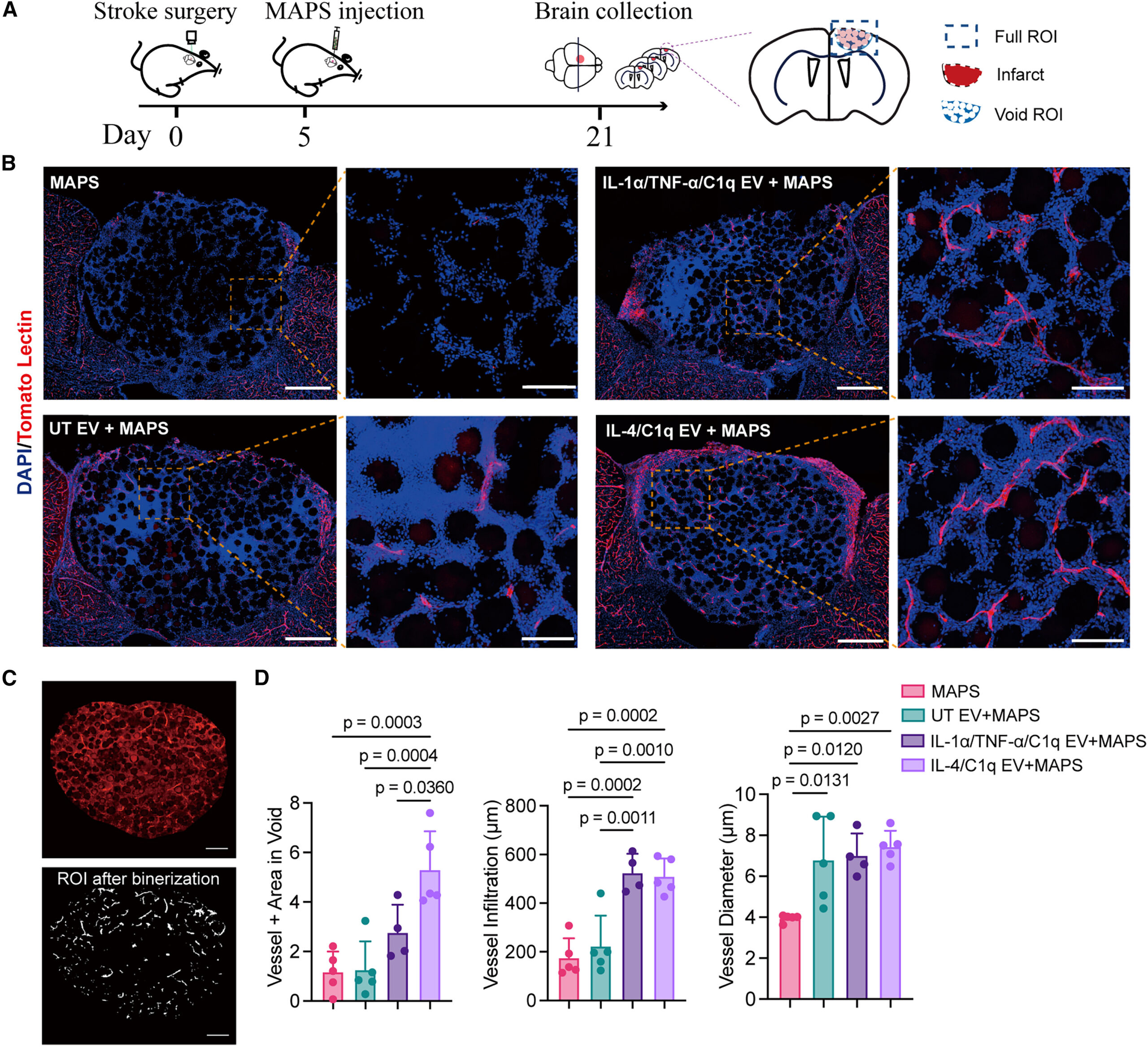
The researchers chemically attached extracellular vesicles directly onto the surfaces of these particles before injecting them into mice five days after stroke, when a well-defined cavity had already formed.
Laboratory testing showed that increasing vesicle loading gradually softened the scaffold because more attachment sites became occupied. The researchers selected a loading level that balanced biological activity with mechanical properties similar to normal brain tissue, while still allowing approximately 20 minutes for injection before gel formation.
One version of the treatment stood apart from all the others
The study compared several groups, including scaffold alone, scaffold carrying untreated astrocyte vesicles, scaffold carrying inflammatory astrocyte vesicles, and scaffold carrying IL-4/C1q astrocyte vesicles.
Only one consistently produced major functional recovery.
Using a standard grid-walking test that measures how often mice miss steps while walking across a wire grid, the researchers found that all stroke-treated animals initially performed poorly.
Mice receiving the IL-4/C1q extracellular vesicle scaffold began improving by 16 days after stroke, while the other treatment groups showed no comparable benefit.
Recovery continued over time.
By about two months, the treated animals’ stepping performance had improved until it became indistinguishable from healthy mice in this behavioral test.
That result suggested something more substantial than short-lived changes in inflammation.
The researchers next examined what had actually happened inside the damaged brain.
New blood vessels rapidly filled the once-empty cavity
One of the most striking observations appeared under the microscope.
Normally, the center of a mature stroke cavity contains almost no functioning blood vessels.
After treatment with the IL-4/C1q vesicle scaffold, however, perfused microvessels extended deep into the damaged region. These vessels were connected to the animals’ circulation rather than simply resembling vascular structures.
Compared with scaffold alone or scaffolds carrying untreated vesicles, the IL-4/C1q treatment generated substantially greater vessel area, deeper penetration into the damaged tissue, and longer vascular branches.
The newly formed vessels were still relatively immature, with incomplete coverage by supporting pericytes compared with healthy brain vessels. Nevertheless, additional analyses showed clear recruitment of these supporting cells, indicating that vascular maturation had begun.
This rebuilding of circulation occurred remarkably quickly, within just over two weeks after implantation.
The treatment also reshaped the brain’s wiring
Blood vessels alone cannot restore brain function.
The researchers therefore examined whether nerve fibers were also returning.
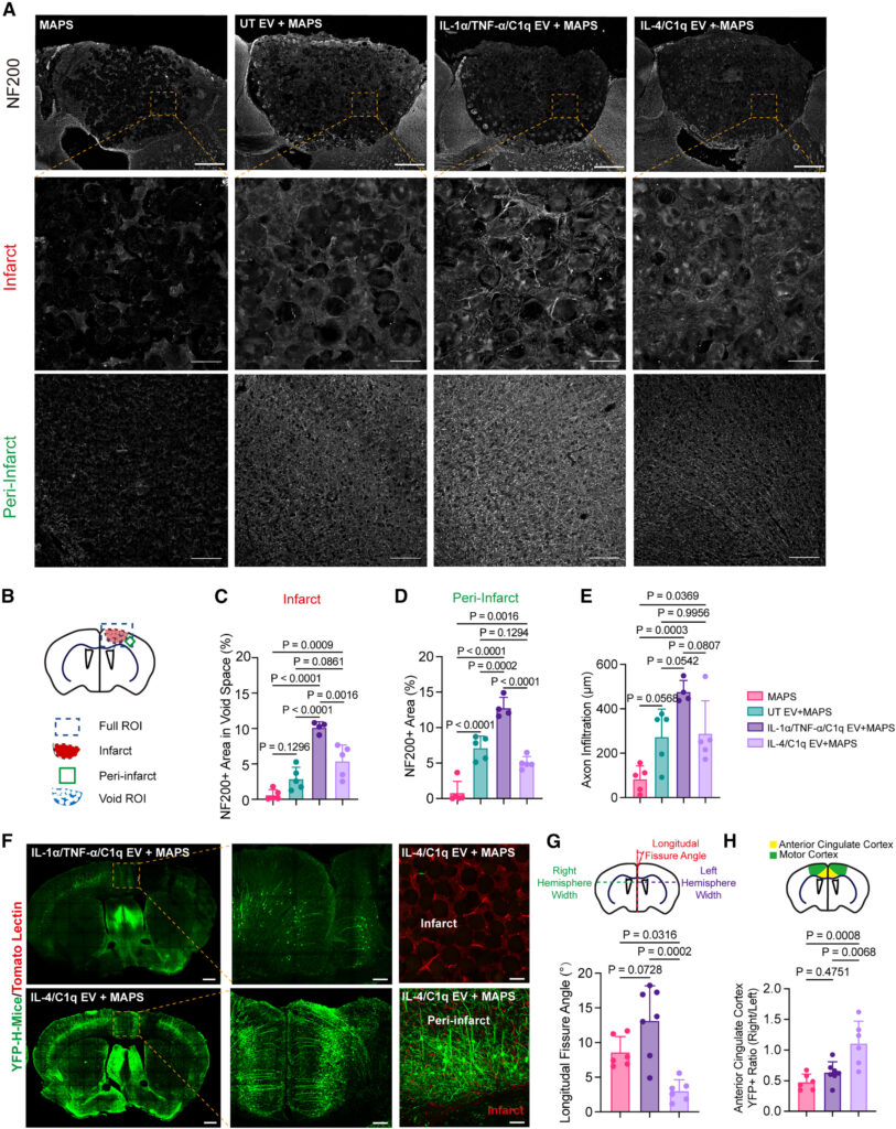
Both activated vesicle treatments increased the growth of axons, the long extensions neurons use to communicate.
However, an important difference emerged.
Although inflammatory astrocyte vesicles produced abundant axonal sprouting, only the IL-4/C1q vesicles promoted rebuilding of nerve fibers associated with motor and sensory pathways in tissue surrounding the stroke.
Experiments using fluorescent reporter mice allowed the researchers to visualize these connections more directly.
The damaged cavity itself still lacked surviving neuronal cell bodies, but surrounding brain regions receiving the IL-4/C1q treatment displayed much stronger ingrowth of labeled nerve fibers.
The treated brains also recovered much of their normal structural symmetry, indicating less long-term distortion after injury.
The findings suggested that simply encouraging axons to grow is not necessarily enough. The pattern of growth—and the biological environment supporting it—appears to matter.
The brain’s repair crew arrived from outside the brain
Perhaps the most surprising part of the study involved immune cells.
Immune cells are often viewed as contributors to stroke damage because many participate in inflammation shortly after blood flow stops.
But the researchers suspected that later in recovery, some of these same cell types might play entirely different roles.
Single-cell RNA sequencing performed nine days after scaffold implantation revealed dramatic changes in the cellular composition of the injured brain.
Compared with scaffold alone, extracellular vesicle treatments attracted many more monocytes, macrophages, and neutrophils into the damaged tissue while reducing the relative abundance of resident glial cells.
Endothelial cells involved in forming blood vessels also became much more common, matching the vascular regrowth seen under the microscope.
Further analyses showed that the IL-4/C1q scaffold particularly favored immune populations linked to tissue remodeling rather than prolonged inflammation.
The researchers also observed changes among T-cell populations, including increases in regulatory T cells and other immune subsets associated with resolving inflammation and supporting vascular remodeling.
Together, these findings suggested that the scaffold was not merely replacing missing tissue. It was creating a biological environment that encouraged the body’s own repair systems to enter and remain within the damaged region.
Neutrophils turned out to be unexpectedly essential
Among all the immune cells entering the scaffold, neutrophils drew particular attention.
Neutrophils have long been associated with early injury after stroke, where they can worsen inflammation and tissue damage.
Yet this study uncovered a very different role later in recovery.
Computational analyses identified neutrophils and macrophages as the immune populations with the strongest expression of genes linked to promoting blood vessel formation.
The IL-4/C1q scaffold recruited the greatest numbers of both.
To determine whether neutrophils were actually necessary, the researchers depleted them using antibodies during recovery.
The consequences were clear.
Without neutrophils, formation of new blood vessels dropped sharply, and remodeling of the hydrogel scaffold slowed substantially.
Rather than acting solely as destructive inflammatory cells, neutrophils appeared to become essential partners in rebuilding damaged tissue during this later stage of healing.
Tiny microRNAs may help explain why one treatment worked
The team next searched for molecular clues explaining why IL-4/C1q-derived vesicles behaved so differently.
Sequencing of vesicle microRNAs revealed a distinctive cargo enriched in the IL-4/C1q group.
Among the molecules attracting attention were miR-377-3p and miR-143-5p, whose predicted target genes were especially active in macrophages and neutrophils.
Previous work has linked these microRNAs with processes including vascular remodeling, neurite growth, and strengthening leukocyte adhesion.
The IL-4/C1q vesicles were also enriched for microRNAs predicted to influence pathways involved in tissue repair, including Wnt signaling, TGF-β signaling, axon guidance, cell adhesion, and related regenerative mechanisms.
Importantly, vesicles produced after exposure to C1q alone failed to reproduce the regenerative effects, indicating that IL-4 played a critical role in programming astrocytes to package these repair-associated molecular messages.
The findings come with important limits
Despite the encouraging results, the researchers emphasize several limitations.
Although neutrophil depletion established an important role for these immune cells, the precise contributions of different macrophage populations remain uncertain.
The extracellular vesicles were isolated using a precipitation method that can also collect non-vesicle material, although all experimental groups were processed identically.
The study was also performed in mice using an experimental stroke model, meaning additional work will be required before any clinical application can be considered.
The researchers say future studies should define exactly how different immune cell populations coordinate repair over time, refine the biological cargo carried by extracellular vesicles, evaluate long-term safety, and eventually test the strategy in larger animal models.
Still, the work challenges one of stroke biology’s longest-standing assumptions.
Instead of viewing the cavity left behind after a stroke as permanently beyond repair, the study suggests it can become an active site of regeneration when supplied with the right biological signals. Rather than replacing lost brain cells directly, the treatment appears to persuade the injured brain—and the immune system beyond it—to rebuild together.
More information
Shangjing Xin et al, IL-4/C1q activated astrocyte-derived extracellular vesicles promote stroke infarct recovery by recruiting peripheral leukocytes, Cell Biomaterials (2026). DOI: 10.1016/j.celbio.2026.100543