Fatty acids produced by gut bacteria activated a nuclear receptor involved in metabolism, while giving vaccenic acid to mice with diet-induced obesity lowered blood glucose and improved several measures of fatty liver disease. The experiments also linked the response to reduced activity of genes involved in making new fat in the liver.
The researchers were looking for a molecular link between gut bacteria and LRH-1, a nuclear receptor that regulates gene activity in tissues including the intestine and liver. Nuclear receptors respond to small molecules and can alter the expression of genes involved in metabolism and other biological processes. LRH-1 had been studied extensively, but the researchers noted that a definitive physiological ligand for the receptor had not yet been identified.
The team began with Bifidobacterium animalis subsp. lactis B420, a probiotic strain whose effects had previously been investigated in metabolic and intestinal models. They extracted lipids from the bacteria and tested whether those molecules could activate LRH-1.
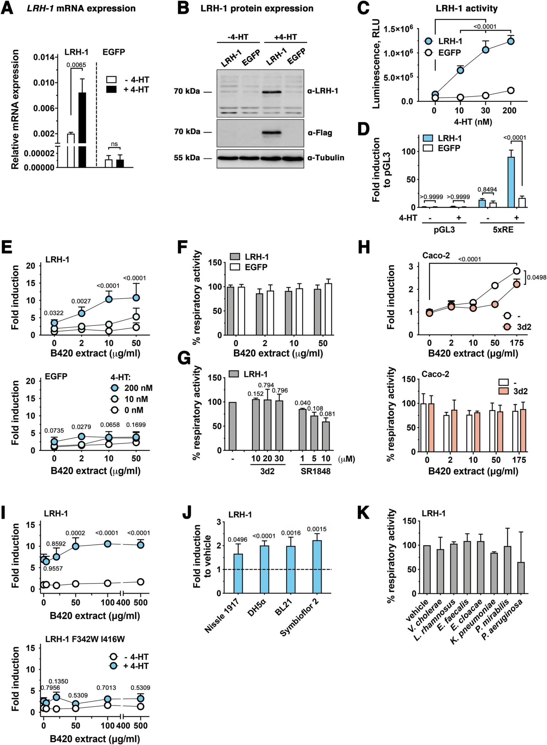
For the initial experiments, the researchers used human LS174T intestinal epithelial cells engineered so that LRH-1 expression could be switched on with 4-hydroxytamoxifen. The cells also carried a luciferase reporter that produces a measurable signal when LRH-1 activates its target DNA sequence.
The B420 lipid extract strongly increased LRH-1 reporter activity at 50 micrograms per milliliter. The response increased with the amount of induced LRH-1 and was also observed in Caco-2 cells, which contain endogenous LRH-1. Two different LRH-1 inhibitors reduced the response. The extract did not produce a corresponding loss of cell viability in the controls.
The investigators then altered two amino acids in LRH-1, producing a mutant receptor designed to interfere with ligand binding in the receptor’s ligand-binding pocket. The mutant showed much less response to the bacterial extract than normal LRH-1, supporting the idea that a molecule in the extract was acting through that pocket.
The phenomenon was not unique to B420. Lipid extracts from several other bacterial strains, including Lactobacillus rhamnosus, Proteus mirabilis, Enterobacter cloacae, Vibrio cholerae, Klebsiella pneumoniae and Enterococcus faecalis, also stimulated LRH-1 activity in the cell assay. Extracts from several Escherichia coli strains did so as well. Pseudomonas aeruginosa was an exception: its extract did not stimulate LRH-1 and instead inhibited reporter activity, an effect the researchers noted could partly reflect reduced cell viability.
The active molecule turned out to be vaccenic acid
Finding an active bacterial extract was only the first step. The researchers needed to determine which molecule was responsible.
They first ruled out DLPC, a synthetic phospholipid known to activate LRH-1. Mass spectrometry did not detect the corresponding DLPC signal in the B420 extract. The team then repeatedly separated the bacterial lipids by chromatography, testing each fraction for LRH-1 activity after every separation.
The most active fraction was purified further by high-performance liquid chromatography. The researchers analyzed the resulting material using several forms of nuclear magnetic resonance spectroscopy and gas chromatography-mass spectrometry. Those analyses identified the active compound as vaccenic acid, or VA, an 18-carbon fatty acid containing one double bond. Both cis and trans forms were present.
The researchers confirmed the presence of the two VA forms in B420 with gas chromatography-mass spectrometry. They also examined other bacterial extracts. Cis-vaccenic acid was detected in all of the other bacterial extracts tested, while trans-vaccenic acid was detected only in B420 among those strains.
When the two purified VA isomers were tested separately, both activated LRH-1 in cells expressing the receptor. The response was dose-dependent, with activity detectable at concentrations between 0.1 and 1 micromolar and reaching its maximum in these experiments at 100 micromolar. The LRH-1 ligand-binding mutant showed a substantially reduced response to both forms.
The effect was also detectable with mouse LRH-1, although the response was weaker than with the human receptor. B420 extracts activated mouse LRH-1, and both VA isomers stimulated its activity.
The fatty acid bound directly to LRH-1
The researchers next asked whether VA was actually binding to LRH-1 rather than merely triggering the receptor indirectly.
They purified the ligand-binding domain of human LRH-1 and measured interactions with VA using microscale thermophoresis, a technique that can estimate how tightly a molecule binds to a protein.
Cis-vaccenic acid bound the LRH-1 ligand-binding domain with a measured dissociation constant of 14.2 micromolar. Trans-vaccenic acid bound more weakly, with a dissociation constant of 144.4 micromolar. Several other long-chain fatty acids tested did not show binding at relevant dissociation constants.
That comparison was important because the bacterial extract contained several fatty acids. The researchers detected palmitic acid, stearic acid and oleic acid among the major lipid species in B420. But oleic acid, elaidic acid, linoleic acid and cis-9,trans-11 conjugated linoleic acid did not activate LRH-1 in the cell-based assay. The researchers suggested that the position or configuration of a fatty acid’s double bond may help determine whether it can interact with the receptor, while also noting that metabolism or instability of the tested compounds could not be excluded as explanations for some of the differences.
The experiments also showed that VA did not activate every nuclear receptor tested. It failed to stimulate PPAR-gamma/RXR or the glucocorticoid receptor in the reported assays, although it did stimulate PPAR-alpha activity, particularly when RXR was also present. The authors therefore described the activity as selective for LRH-1 among the receptors examined, rather than as an effect unique to LRH-1 among all possible cellular targets.
One difference between the two VA isomers remained unresolved. Trans-VA bound LRH-1 about ten times more weakly than cis-VA in the protein-binding experiment, yet both produced similar activation in the cell assay. The authors proposed that factors inside cells, including interactions with cofactors, metabolic conversion, nonlinear responses or differences in bioavailability, could help explain the discrepancy. They did not establish which explanation was responsible.
VA changed LRH-1-controlled genes in mice
The next experiments moved from isolated cells to animals.
Male C57BL/6 mice received an intraperitoneal injection containing a mixture of cis- and trans-VA at 50 milligrams per kilogram. Tissue was collected eight or 18 hours later.
In the small intestine, VA increased expression of Cyp11a1 and Cyp11b1, genes regulated by LRH-1 and involved in intestinal steroid production. When the researchers isolated intestinal crypt cells from mice lacking LRH-1 specifically in intestinal epithelial cells, the VA-induced response was not reproduced under the tested conditions, although the difference for Cyp11a1 did not reach statistical significance.
VA also altered gene expression in the liver. After a single injection, expression of Scd1, Cyp7a1 and Cyp8b1 decreased. Serum glucose was also significantly lower after the injection, while serum insulin did not change significantly in that experiment, which involved three mice per condition.
The researchers additionally observed increased expression of Shp in the liver and Fgf15 in the intestine. Because those genes are associated with the FXR-SHP-FGF15 pathway, the authors suggested that VA’s effects could involve interactions between LRH-1 and FXR signaling. The experiments did not establish that this pathway was responsible for the metabolic effects.
The strongest metabolic effects appeared in obese mice
To test whether the receptor response was accompanied by changes in metabolic disease, the researchers used male mice fed either a standard diet or a high-fat diet for 15 weeks.
The high-fat diet produced the expected metabolic phenotype in this experiment. Compared with standard-diet mice, the high-fat-diet animals had greater body weight and higher blood glucose, serum cholesterol, triglycerides and GPT/ALT, a liver enzyme used here as a measure associated with liver injury. They also had altered liver weight, more liver triglycerides and increased lipid deposition in liver tissue.
After those 15 weeks, the mice received daily intraperitoneal injections of either the cis/trans VA mixture at 50 milligrams per kilogram or vehicle for two weeks while remaining on their assigned diets.
VA did not significantly alter body weight or weight gain during treatment. Nor did the treatment produce evidence of liver toxicity in the measures reported: serum GPT/ALT and normalized liver weight remained comparable with controls.
Blood glucose was measured before treatment and again after seven and 13 days. In the high-fat-diet group, VA treatment lowered blood glucose at both time points. The difference at day 13 approached but did not reach conventional statistical significance, with P = 0.058. Glucose at the end of the experiment was also lower in VA-treated high-fat-diet mice. Serum insulin was lower in the VA-treated high-fat-diet group, while the same effect was not observed in standard-diet mice.
The liver showed a more mixed response. Histological examination indicated a mild reduction in lipid deposition after VA treatment, but total liver triglycerides did not differ significantly. A more detailed assessment separated macrovesicular steatosis, microvesicular steatosis and hepatocyte hypertrophy and combined them into a NAFLD activity score. That score was significantly improved in VA-treated high-fat-diet mice, with the reduction driven mainly by lower microvesicular steatosis and hepatocyte hypertrophy.
VA did not significantly change collagen deposition or fibrosis. It also did not significantly alter serum cholesterol or serum triglycerides.
Liver gene activity pointed to reduced fat production
The researchers then used RNA sequencing to examine liver samples from the VA-treated and vehicle-treated high-fat-diet mice.
The genes that changed significantly after VA treatment were enriched in pathways associated with lipid biosynthesis and fatty acid metabolism. Several genes involved in making new fat were reduced, including Acacb, Fasn and Scd1. Expression of Mid1ip1, which promotes fatty acid biosynthesis through the nuclear receptor LXR, and Cd36, which encodes a fatty acid transporter, also decreased.
Two established LRH-1 targets, Cyp7a1 and Cyp8b1, were also reduced. Independent RT-qPCR measurements confirmed the decrease in Cyp8b1 and Scd1. The reduction in Scd1 was observed in the high-fat-diet setting, while the decrease in Cyp8b1 was also detected in standard-diet mice.
The authors interpreted the combined gene-expression results as evidence that reduced liver lipogenesis could contribute to the glucose and lipid changes seen after VA treatment. That interpretation connects the receptor experiments with the metabolic phenotype, but the study did not establish that suppression of lipogenesis alone caused the improvements.
Several questions remain about where VA acts
The experiments establish several links in the proposed pathway. Lipid extracts from multiple bacteria activated LRH-1 in cell assays. VA was isolated from an active bacterial extract, both VA isomers activated LRH-1, and both bound the purified human LRH-1 ligand-binding domain. VA also changed LRH-1-associated gene expression in mice and altered metabolic measurements in mice with diet-induced obesity and hyperglycemia.
But the experiments do not demonstrate that bacterial production of VA is responsible for the metabolic effects observed in the treated mice. The animals were injected with VA rather than given a VA-producing bacterium, and the study did not directly establish the amount of bacterial VA that normally reaches the liver or whether endogenous bacterial VA activates LRH-1 under physiological conditions.
The authors also note that VA is present in human tissues and plasma and that trans-VA concentrations in human blood have been reported in the micromolar range. They discuss meat and dairy consumption as a major source of VA in humans while also identifying bacteria as another source. Those observations place VA in a physiological context, but they do not demonstrate that dietary or microbiota-derived VA activates LRH-1 in humans.
Another unresolved issue is whether VA’s effects in living animals result entirely from direct LRH-1 activation. The authors explicitly note that VA might instead, or additionally, stimulate the production of other LRH-1 ligands. The study also does not determine why the two VA isomers have markedly different binding affinities but similar effects in cell-based assays.
The study therefore traces a biochemical pathway from bacterial lipids to a host nuclear receptor and then to metabolic changes in mice, while leaving the physiological contribution of that pathway to normal microbiota-host communication unresolved. The RNA-sequencing data and the animal experiments point specifically to altered expression of genes involved in hepatic lipid metabolism as part of the response to VA.
The study was published in EMBO Molecular Medicine.