New photon-counting CT scans are allowing researchers to examine Egyptian mummy remains more than 2,300 years old in much greater detail, including their bones, teeth and bandages, while leaving the remains intact.
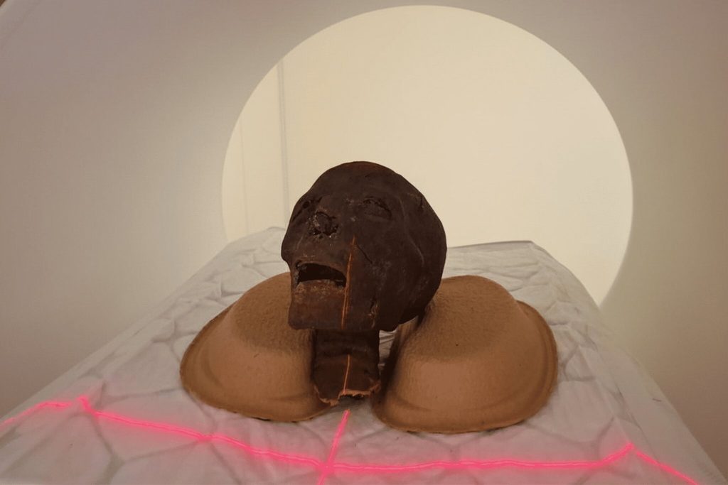
The examinations are being carried out at Semmelweis University’s Medical Imaging Center, where researchers are using a CT scanner equipped with a photon-counting detector. The Egyptian remains came from the Semmelweis Museum of Medical History, part of the Hungarian National Museum Public Collection Center.
The museum’s Egyptian remains have been examined several times in recent years, including with conventional CT scans. But those earlier images were not detailed enough for some questions to be answered.
Radiocarbon dating was attempted on six specimens, although only three produced measurable results. The oldest dated remains fall between 401 and 259 BCE, making them more than 2,300 years old.
Researchers are now reexamining that specimen along with the museum’s other Egyptian mummy remains using the newer imaging system.
The goal is to learn as much as possible about the internal structure of the remains, possible abnormalities and the techniques used to preserve them, said Dr. Ibolyka Dudás, chief clinical physician in the Department of Radiology and head of the post-mortem imaging working group.
The new images provide more detail
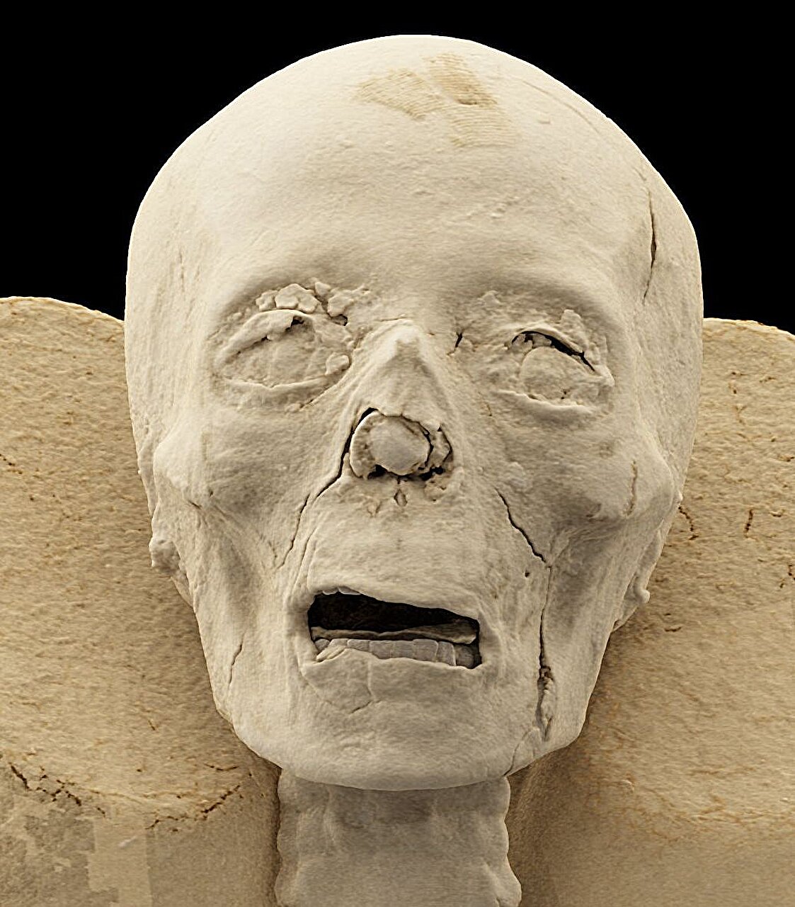
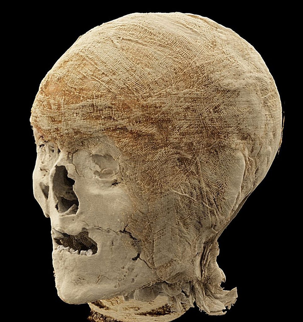
Two mummified heads can now be examined in greater detail, particularly their teeth and the sutures between the skull bones. Those features could help researchers determine age more accurately. The scans may also provide a basis for future high-precision 3D reconstructions and possible facial reconstructions.
A previously examined left lower limb also looks different under the new imaging. Earlier examinations could not establish a definitive diagnosis. The latest images have produced several possible interpretations, including the possibility that the individual had osteoporosis.
Researchers do not yet know whether that condition was related to aging or resulted from a pathological process. More detailed analysis is needed.
Another left lower limb appears to have belonged to a young individual, although the person’s exact age is still being determined. The new scans provide the first detailed imaging data available for that specimen.
A supposed mummy bundle turned out to be a foot
One of the more unusual cases began with an object that was initially classified simply as a mummy bundle.
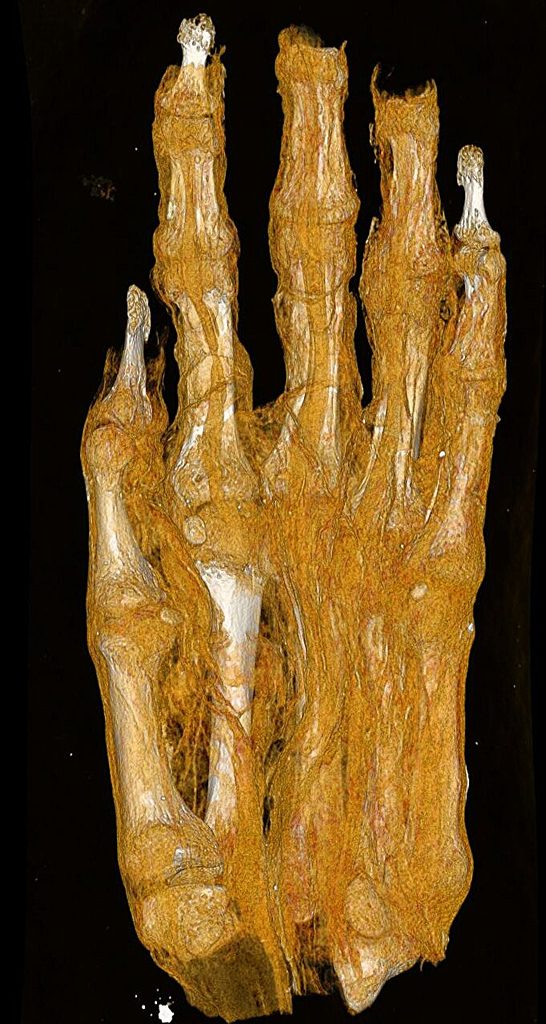
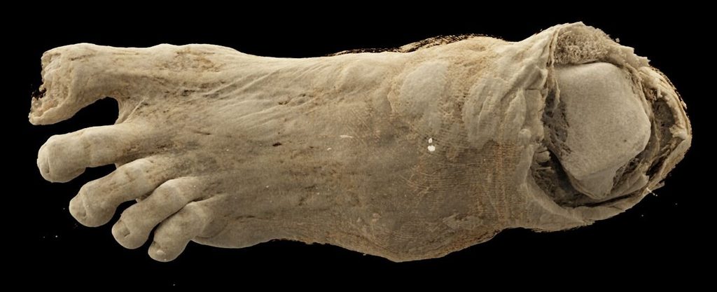
When it reached the museum, it was first thought to be a human head. Without imaging, it was later considered possibly to be a bird mummy. A previous CT scan established that the object was actually an adult foot.
The latest scans are being used to examine the textile remains wrapped around it. Researchers want to determine what the layers can reveal about the mummification technique, the age of the person and possible illnesses.
The new images clearly show multiple layers of bandaging with different structural characteristics. Researchers say this could support further historical and technological investigations.
The foot was presumably part of a complete mummy, but researchers do not yet know why or when it was separated from the rest of the body.
Bones may help identify age and sex
The study also includes a mummified hand. Researchers are examining the size, development and shape of its bones to determine whether it belonged to a child or an adult. Those characteristics may also help estimate the person’s sex and age.
The detailed assessment of all the new images is still underway. Researchers expect the analysis to provide additional information about the individuals’ lives and health, as well as the mummification process.